About Chilean Wool
Chilean Wool, LLC is a data analytics company specializing in analysis of whole slide tissue image data. It was founded by a team with decades of experience in digital pathology, big data analytics, biomedical informatics, and medicine and passion for innovation and transformative technologies.
In the late 90s, realizing the tremendous potential of digitized tissue slides in research, clinical environments and education, we developed Virtual Microscope, one of the first digital pathology software systems. Virtual Microscope enabled remote viewing of whole slide tissue images and paved the way for digital dynamic telepathology. Since then we have researched and implemented innovative solutions in management and analysis of digital pathology data to maximize its value in research and healthcare delivery.
Our recent work led to the first quantitative analysis of lymphocyte patterns in tissue images from multiple cancer types using AI methods. Later work has refined this initial AI methodology and extended it to enable characterizations of spatial relationships between lymphocytes and tumor regions. With increasing evidence of the relationship between immune response to cancer and favorable clinical outcomes, quantitative classifications of tumor infiltrating lymphocytes have great potential to improve prediction of response to treatment and help with selection of best treatment strategies.
Meet the Team
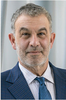
Joel Saltz
Co-founder and Chief Executive Officer
Joel H. Saltz, MD, PhD, is a physician–computer scientist whose career bridges high-performance computing, biomedical informatics, and pathology. He earned his bachelor’s degree in mathematics and physics and master’s degree in mathematics at the University of Michigan, followed by an MD and PhD in computer science at Duke University. He completed his clinical pathology residency at The Johns Hopkins Hospital and is board-certified in clinical pathology and clinical informatics. His computer science research has advanced parallel computing, large-scale data systems, and spatial analytics—providing a foundation for making complex biomedical images and clinical data useful for research and medicine. Dr. Saltz founded and chaired three biomedical informatics departments: at The Ohio State University, Emory University, and Stony Brook University. He also served as Vice Chair of Pathology at both Ohio State and Stony Brook, and held computer science faculty positions at Yale and the University of Maryland. Across these institutions, he built interdisciplinary programs connecting computing, laboratory medicine, cancer research, and clinical care. His digital pathology work dates to the Virtual Microscope, reported by his group in 1997 as the first published system for whole-slide viewing, and extends through scalable image-analysis platforms, reusable pathology AI software, and modern foundation-model and generative AI methods. For the past decade, Dr. Saltz has led a sustained research effort in pathology AI focused on tumors and tumor-infiltrating lymphocytes (TILs), the immune cells found within and around cancers. His group and collaborators developed methods to map these cells across whole-slide tissue images and study how their spatial organization relates to tumor biology, molecular features, and patient outcomes. This publication record includes the 2018 Cell Reports pan-cancer TIL study, subsequent breast cancer spatial-analysis studies, open pathology AI tools such as WSInfer, and recent foundation-model research on breast cancer recurrence risk. He led the pathology analyses in the TCGA tumor/TIL work and was a member of the TCGA team recognized with the 2020 AACR Team Science Award. This combination of computational expertise, pathology training, and tumor–immune research informs his work at Chilean Wool, developing tools to make spatial immune analysis accessible and useful for cancer research.

Rajarsi Gupta, MD, PhD, is a pathologist and informatician with over 20 years of experience. His research focuses on developing Pathomics AI methodologies to detect, quantify, and extract clinically significant features from high-resolution digital whole slide images (WSIs) in cancer histopathology. His work aims to produce practical, reliable, efficient, and cost-effective solutions for translational clinical and biomedical applications. His expertise in Pathomics AI emphasizes mapping tumor-infiltrating lymphocytes (TILs) within the tumor microenvironment to characterize spatial distribution and abundance, supporting precision oncology by predicting clinical outcomes—such as survival, disease progression, molecular profiles, and treatment response—and guiding personalized immunotherapy selection. Over the past eight years, he has co-led initiatives to design, develop, validate, implement, and refine machine learning and artificial intelligence approaches in computer vision for analyzing diverse histologic cancer specimens.
Rajarsi Gupta
Co-founder and Chief Scientific Officer

Mary Saltz, MD, is a diagnostic radiologist and board-certified clinical informatician with over 30 years of experience. Her clinical informatics training encompasses human-computer interaction, user interface design, clinical workflow optimization, information display, and usability. She has held leadership positions as a radiology department chair, Chief Quality Officer, Clinical Information Officer of the Stony Brook Cancer Center, and Chief Integration Officer. Her informatics research has centered on clinician interactions with complex medical data, resulting in the development of a serverless browser system for real-time analysis of 20 million New York State SPARCS records, platforms for generating and exploring whole-slide image features, multi-class medical image segmentation, and integrated radiomics and pathomics datasets.
Mary Saltz
Co-founder and Chief Medical Officer

Tahsin Kurc
Co-founder and Chief Technology Officer
Tahsin Kurc, PhD, is a computer scientist with over 30 years of expertise in high-performance computing, biomedical informatics, and digital pathology. His research centers on developing scalable computational methods and software systems for the management, analysis, and interpretation of large biomedical datasets, with a primary focus on whole-slide tissue imaging. Dr. Kurc’s early work addressed foundational challenges in high-performance data management and scientific computing, contributing to pioneering systems that introduced scalable strategies for accessing, processing, and visualizing expansive scientific and biomedical data. He subsequently developed high-performance computing techniques that enabled efficient image-analysis workflows across multi-core processors, hardware accelerators, and distributed-memory systems for terabyte-scale datasets. Building on this foundation, Dr. Kurc helped advance informatics infrastructures for digital pathology, incorporating database, service-oriented, web-based, and cloud technologies to manage, analyze, visualize, and share large collections of whole-slide images and associated analytical data. His current work emphasizes artificial intelligence and computational pathology, including deep learning for tissue segmentation and classification, pathomics, spatial characterization of tumor regions and tumor-infiltrating lymphocytes (TILs), and computational biomarkers linked to clinical outcomes. Recent investigations also examine advanced representation learning and generative approaches in histopathology, extending his long-standing contributions at the intersection of scalable computing, biomedical imaging, and AI.
Selected publications from our team
Spatial Organization and Molecular Correlation of Tumor-Infiltrating Lymphocytes Using Deep Learning on Pathology Images (Link to Full Article)
Digitized H&E-stained images of TCGA samples, beyond sample curation and basic pathologic characterization, remain underutilized. This work presents mappings of tumor-infiltrating lymphocytes (TILs) based on H&E images from 13 TCGA tumor types. These TIL maps are derived through computational staining using a convolutional neural network trained to classify image patches. Affinity propagation revealed local spatial structure in TIL patterns and correlation with overall survival. TIL map structural patterns were grouped using histopathological parameters and enriched in particular T cell subpopulations derived from molecular measures. TIL densities and spatial structure were differentially enriched among tumor types, immune subtypes, and tumor molecular subtypes, implying that spatial infiltrate state reflects particular tumor cell aberration states. Obtaining spatial lymphocytic patterns linked to the rich genomic characterization of TCGA samples provides insights into the tumor-immune microenvironment.
Towards interpretable prediction of recurrence risk in breast cancer using pathology foundation models (Link to Full Article)
Transcriptomic assays like the PAM50-based ROR-P score guide recurrence risk stratification in non-metastatic, ER-positive, HER2-negative breast cancer. However, these assays are not universally accessible. Histopathology, a routinely available alternative, offers a scalable solution. In this work, foundation models, large neural networks pre-trained on pathology images, were trained and validated on the Carolina Breast Cancer Study and externally tested on TCGA BRCA. Several foundation models outperformed baseline models across classification, regression, and survival tasks. CONCH achieved the highest ROC AUC, while H-optimus-0 and Virchow2 showed the highest correlation with continuous ROR-P scores. All pathology models stratified CALGB participants by recurrence similarly to transcriptomic ROR-P. Tumor regions were necessary and sufficient for high-risk predictions and identified candidate tissue biomarkers of recurrence. These results highlight the promise of interpretable, histology-based risk models in precision oncology.
Mapping tumor infiltrating lymphocytes in whole prostatectomy specimens to visualize and quantify the immune microenvironment of prostate cancer (Link to Full Article)
Prostate cancer (PCa) frequently presents as multifocal lesions with histologic and molecular heterogeneity. A comprehensive understanding of tumor immune responses within the context of the complex PCa microenvironment remains elusive. Through the use of Digital Pathology and innovative Pathomics image analysis tools, tumor infiltrating lymphocytes (TILs) can be mapped, visualized, and quantified in whole slide images (WSIs) of H&E stained PCa slides. We have developed a novel workflow to map and quantify TILs by Gleason histologic grade (Gleason-TIL maps) in whole radical prostatectomy specimens.
Spatial Characterization of Tumor-Infiltrating Lymphocytes and Breast Cancer Progression (Link to Full Article)
Tumor-infiltrating lymphocytes (TILs) are a robust prognostic biomarker in breast cancer, predicting treatment response in both adjuvant and neoadjuvant settings. This study evaluated TILs’ role in predicting overall survival and progression-free interval in two independent breast cancer cohorts from the TCGA BRCA and UNC CBCS. We used machine learning and computer vision algorithms to characterize TIL infiltrates in digital H&E-stained WSIs. Global abundance and spatial features of TILs were analyzed. Univariate and multivariate analyses showed that large peritumoral and intratumoral TIL aggregates (forests) correlated with longer survival, while intratumoral TIL absence (deserts) correlated with increased recurrence risk. Patients with two or more high-risk spatial features had significantly shorter progression-free interval. This study demonstrates Pathomics’ utility in evaluating TIL abundance and spatial patterns as important biomarkers in breast cancer.
Deep Learning-Based Mapping of Tumor Infiltrating Lymphocytes in Whole Slide Images of 23 Types of Cancer (Link to Full Article)
This study presents an updated deep learning workflow to classify 50x50 um tiled image patches (100x100 pixels at 20x magnification) as TIL positive or negative based on the presence of two or more TILs in gigapixel whole slide images (WSIs) from the Cancer Genome Atlas (TCGA). This workflow generates TIL maps to study TIL abundance and spatial distribution in 23 cancer types. Our study trained three state-of-the-art convolutional neural network (CNN) architectures (VGG16, Inception-V4, and ResNet-34) with a large training dataset. The dataset includes TIL positive and negative patches from cancers in additional organ sites and curated data to enhance algorithmic performance. Automated thresholding converts model predictions into binary classifications to generate TIL maps. The new TIL models achieve superior performance, with improvements of up to 13% in accuracy and 15% in F-score.
Utilizing Automated Breast Cancer Detection to Identify Spatial Distributions of Tumor-Infiltrating Lymphocytes in Invasive Breast Cancer (Link to Full Article)
This study developed and evaluated CNN analysis pipelines to generate combined maps of cancer regions and TILs within routine diagnostic breast cancer whole slide tissue images. These maps provide insights into lymphocytic infiltrates’ structural patterns and spatial distribution, enhancing TIL quantification.